
Optimizing Mammography Performance Through Advanced QA Technology
As mammography continues to evolve, new detector technology is redefining how kVp, HVL, dose, and other key parameters are measured with accuracy and efficiency.
Precision Matters: Elevating QA in Mammography
Mammography screening is uniquely demanding within diagnostic imaging since most of the patients are healthy with no disease in the exposed area. Designed to detect the earliest signs of breast cancer, it relies on exceptional contrast sensitivity to distinguish subtle variations in soft tissue. At the same time, it operates at relatively low X-ray energies, where even small inconsistencies can affect both image quality and patient dose.
This makes quality assurance (QA) not just a regulatory requirement, but a cornerstone of clinical confidence.
Unlike general radiography, mammography systems use a wide variety of target and filter combinations to shape the X-ray spectrum. From standard screening to contrast-enhanced techniques, beam qualities can vary significantly, typically spanning 18 to 49 kV. Ensuring consistent system performance across this range is one of the key challenges facing medical physicists and service engineers.
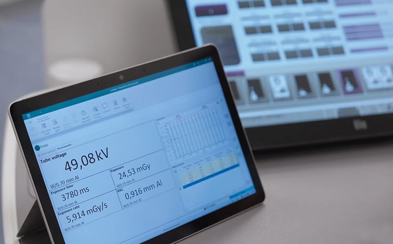
Picture 1. With the Ocean computer software all measured parameters can be seen at the same time, including waveform.
Measuring What Matters
Effective QA in mammography focuses on verifying both radiation output and beam quality. Core measurements include:
• Tube voltage (kVp)
• Dose and dose rate
• Exposure time
• Half-value layer (HVL)
These parameters directly influence image contrast and patient exposure. They also form the basis for calculating Average Glandular Dose (AGD), widely recognized as the most relevant dose metric in breast imaging.
Given the clinical importance of these values, accuracy and traceability are essential. International protocols such as ACR and EUREF place strong emphasis on reliable measurement methods and repeatable workflows.
Mako: Built for Mammography
RTI Group’s Mako system introduces a new level of capability for mammography QA through its dedicated Mako Mammo Probe.
A defining advantage is its ability to cover the entire clinical kV range (18–49 kV) without requiring multiple detectors. This allows users to perform both standard and advanced QA tasks, with a single setup across all measurement parameters.
The Mako Mammo Probe advanced detector covers all existing mammography beam qualities on the X-ray Mammography market, covers older systems and the newest additions to the market, including emerging target/filter combinations such as titanium. See picture 2.
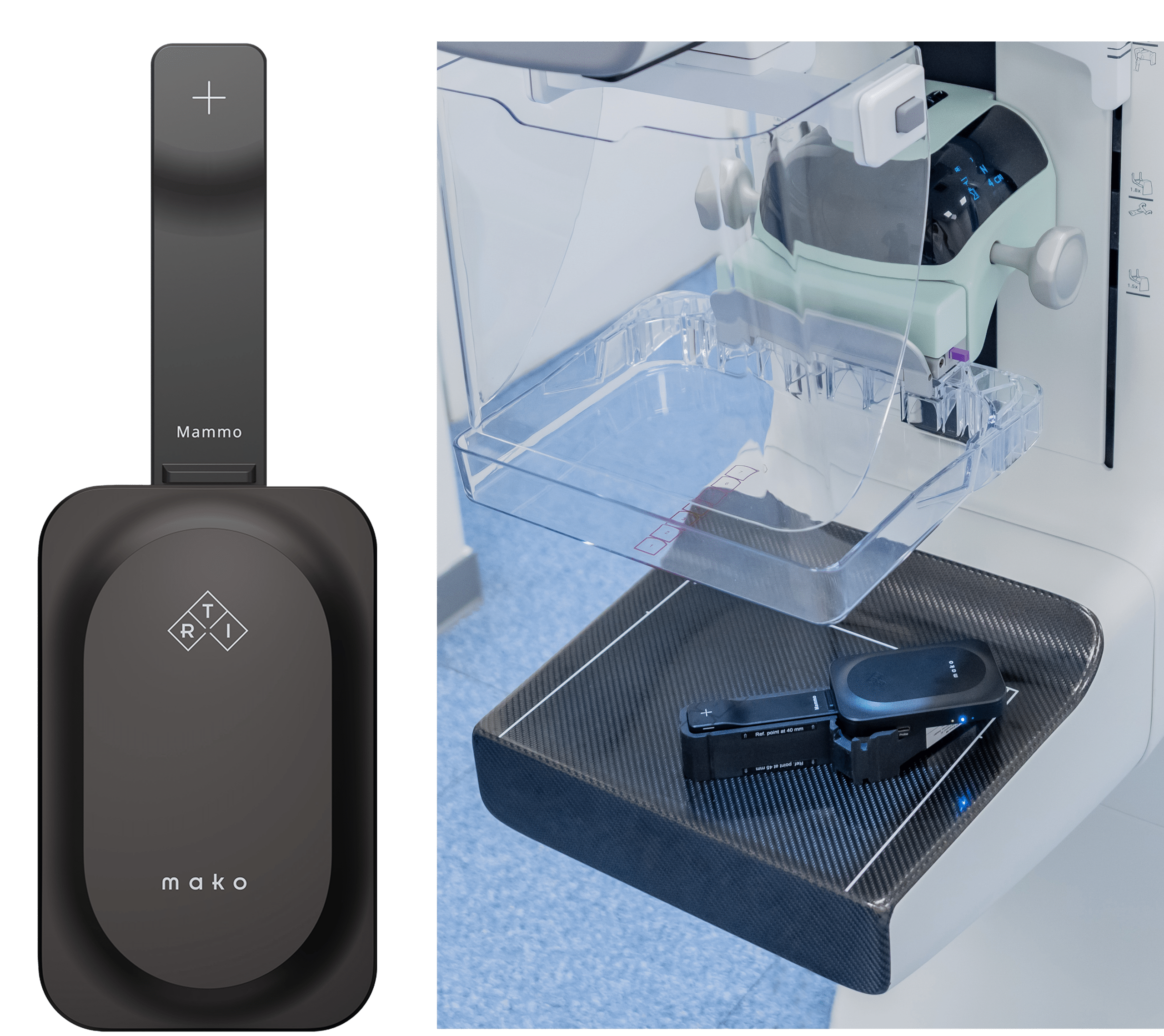
Picture 2, a) Mako Base Unit with Mako Mammo Probe, b) Mako Mammo Holder
Reducing Complexity in Daily QA
In practice, QA efficiency depends as much on usability as on accuracy. Mammography setups can raise practical questions: positioning relative to the chest wall, handling of the compression paddle, or adapting to different system geometries.
Mako addresses these challenges through intelligent design:
- Simply place and measure with the Mako Probe, no orientation-dependence. Reduces setup errors and makes Mako robust across systems and users
- No need to worry about the compression paddle. Mako has automatic compensation to reduce the user selections required
- No selections required to measure at all with Mako. The user always has the option just measure dose, HVL and time without any manual input
In some markets the meter should be at 40 mm or 45 mm hight. The RTI Mako Mammo Holder enables easy, precise positioning of the Mako Mammo Probe at both these heights, see Picture 2 b.
Supporting Advanced Imaging: Tomosynthesis
The increasing adoption of digital breast tomosynthesis (DBT) adds another layer of complexity. As the X-ray tube moves through multiple angles, maintaining accurate dose measurement becomes more difficult.
Mako’s detector enables reliable measurements across wide angular ranges, supporting QA in both 2D and 3D imaging without repositioning for ±30° angle.
The Role of the Mako Display
Designed as a fully wireless, app-powered interface, the Mako Display removes the need for cables, external laptops, or complex setup. Measurements are streamed in real time directly to the user’s device, providing immediate access to kVp, dose, HVL, waveforms, and other key parameters at the point of testing.
The app is available as a free download with no licenses or user restrictions, making it easy to deploy across teams and devices without additional cost or complexity.
Mako is also built for flexibility. When needed, the Mako Probe can be connected to the Base Unit via cable, to allow more delicate positioning. Mako can also connect to a full fleet of external detectors like mAs meters, light meters and ion chamber detectors. And when deeper QA work is needed with analysis, calculation and reporting, the RTI computer software Ocean Next can be used to automate QA routines.
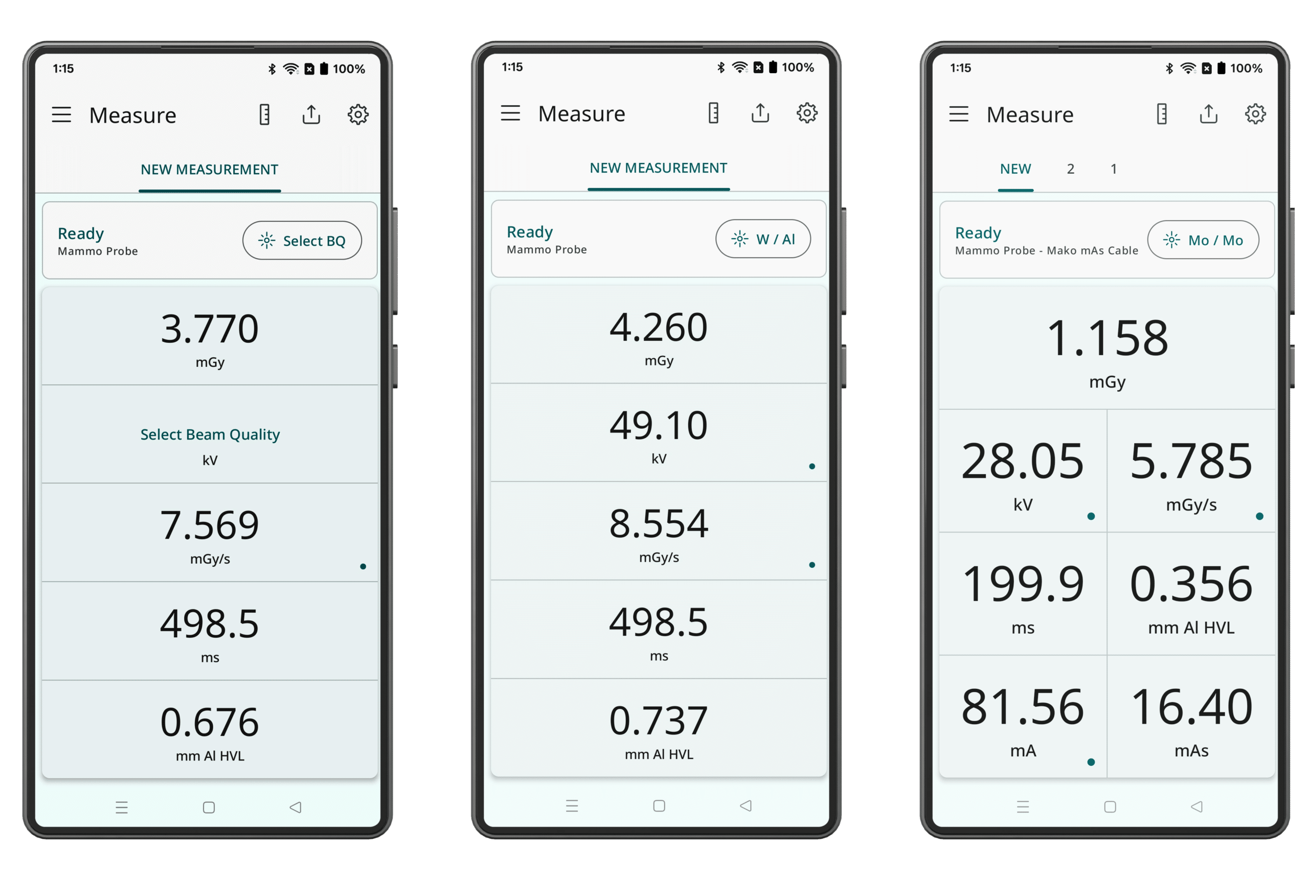
Mako Display a) No selected Beam Quality, b) Selected Beam Quality c) Mako Mammo Probe with mAs-meter connected (simultaneous tube & generator measurement)
More Reliable Mammography QA
As breast imaging technology evolves, QA must keep pace with increasing complexity and higher expectations. The combination of accurate measurement, simplified setup, and integrated software tools represents a more sustainable approach to quality assurance.
With Mako and its connected display environment, QA becomes not only more precise, but also more efficient and easier to manage. The result is a workflow that supports clinicians in delivering high-quality mammograms with confidence—every time.

Björn Cederquist is a Sweden-based medical physicist who joined RTI Group in 2008. Starting in R&D with detector design, he moved into sales and technical support in 2009. Today, he supports customers across mainland Europe, Japan, and South Korea, combining strong technical expertise with practical clinical insight.
Published in EFOMP News, Issue 02/2026/Summer, pages 58-58 and 81.